THIS PRODUCT IS DISCONTINUED
Fast Activated Cell-based ELISA (FACE™) Kits provide a simple, sensitive method for detecting protein phosphorylation directly in the cell, without making extracts or performing electrophoresis and membrane blotting. These 96-well, high-throughput assays are available in both colorimetric and chemiluminescent formats for over 20 different targets (see list at right). For complete details, click the FACE™ Method tab below.
Each FACE ErbB-2 Kit provides 96 rxns each of 2 antibodies that enable you to monitor and compare the levels of both phosphorylated and total ErbB-2. The Y877 kit contains phospho-ErbB-2 antibody that recognizes ErbB-2 only when phosphorylated at Tyr877, while the Y1248 kit provides a phospho-antibody that recognizes ErbB-2 only when phosphorylated at Tyr1248. Each kit contains a total-ErbB-2 antibody that recognizes ErbB-2 regardless of its phosphorylation state. Click the ErbB-2 Info tab below for data and more information.
| Name | Format | Cat No. | Price | |
|---|---|---|---|---|
| FACE™ ErbB-2 (Y877) | 1 x 96 rxns | 48130 | Discontinued | |
| 5 x 96 rxns | 48630 | Discontinued | ||
| FACE™ ErbB-2 (Y877) Chemi | 1 x 96 rxns | 48230 | Discontinued | |
| 5 x 96 rxns | 48730 | Discontinued | ||
| FACE™ ErbB-2 (Y1248) | 1 x 96 rxns | 48105 | Discontinued | |
| 5 x 96 rxns | 48605 | Discontinued | ||
| FACE™ ErbB-2 (Y1248) Chemi | 1 x 96 rxns | 48205 | Discontinued | |
| 5 x 96 rxns | 48705 | Discontinued | ||
| FACE™ ErbB-2 Manual |
| FACE™ Profile |
| Cell Biology Products Brochure |
| IsoCyte™ Application Note – Phospho-Protein Detection |
| MSDS: Sodium Azide |
| MSDS: Sulphuric Acid |
| MSDS: Thimersol |

Figure 1: Monitoring total- and phospho-ErbB-2 using FACE.The FACE ErbB-2 (Y1248) and ErbB-2 (Y877) Kits were used to assay the levels of total and phosphorylated ErbB-2 contained within untreated or EGF treated A-431 cells.
Antibody Specificities
The FACE ErbB-2 (Y877) Kit contains a phospho-ErbB-2 antibody that was raised in rabbit against a synthetic phospho-peptide corresponding to residues surrounding phosphorylated Tyr877 of human ErbB-2 and recognizes ErbB-2 only when phosphorylated at Tyr877. The FACE ErbB-2 (Y1248) Kit contains a phospho-ErbB-2 antibody that was raised in rabbit against a synthetic phospho-peptide corresponding to residues surrounding phosphorylated Tyr1248 of human ErbB-2 and recognizes ErbB-2 only when phosphorylated at Tyr1248. The total-ErbB-2 antibody supplied in the FACE ErbB-2 Kits recognizes ErbB-2 protein regardless of its phosphorylation site.
ErbB-2 Overview
The ErbB-2 proto-oncogene, also called Neu, EGFR-2 or HER-2, is a member of the transmembrane receptor tyrosine kinase family, which also includes EGF receptor and EGFR-3 (HER-3 or ErbB-3). Although, ErbB-2 does not have any known high-affinity ligands, its kinase activity can be activated without ligand by either overexpression or heteroassociation with other members of the ErbB family. Amplification of the ErbB-2 gene and overexpression of its product has been detected in almost 40% of primary human breast tumors, which correlates with poor prognosis in node positive breast cancer. ErbB-2 overexpression is also observed in ovarian, gastric, salivary and non-small cell lung carcinomas. Because of this, ErbB-2 is one of the major drug targets for breast cancer and other cancer treatments.
FACE™ の使用法
96ウェルプレートで細胞を培養し, 目的の経路を誘導する刺激を与えます。刺激を与えた後, 細胞を固定することでリン酸化を含むタンパク質修飾が保持されます。ブロッキング反応の後, それぞれのウェルに目的タンパク質に特異的な一次抗体, 続いてHRP標識二次抗体を加えます。その後, 発色試薬を加えることにより, 比色定量または化学発光による定量を簡便に行うことができます (図1)。また, キット付属のCrystal Violetを用いて細胞数を補正することができます。それぞれのFACE Kitには目的タンパク質全体とリン酸化型を認識する一次抗体が含まれます。これにより, 細胞数と細胞中の目的タンパク質の総量に対してのリン酸化型タンパク質量を解析することができます。FACEはリン酸化型タンパク質を解析することのできるキットで, 定量的かつ再現性のある結果が得られます。
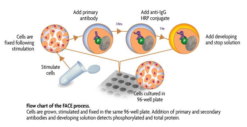
図1: FACE の操作法
Contents & Storage
Two (or ten) 96-well plates for culturing cells, 96 (or 5 x 96) rxns each of two primary antibodies (1 phospho-specific, 1 specific for native protein), HRP-conjugated secondary antibody, Quenching Solution, 1X Antibody Blocking Buffer, 1X Antibody Dilution Buffer, 10X PBS, 10% Triton X-100, 1% SDS Solution, Developing and Stop Solutions, and Crystal Violet Cell Quantification Solution. Storage conditions vary from room temperature to -20°C, see manual for details. All reagents are guaranteed stable for 6 months when stored properly.