<< Back to MOTIFvations Blog Home Page
Hypoxia Researchers Win 2019 Nobel Prize in Physiology or Medicine
By Anne-Sophie Ay-Berthomieu, Ph.D.
November 8, 2019
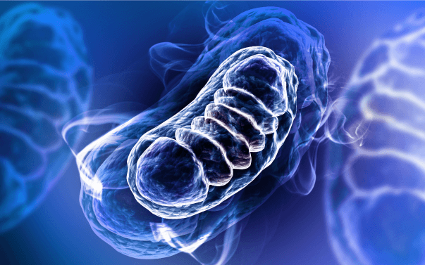
Energy production in multicellular organisms is carried out by the mitochondria and relies on the presence of molecular oxygen (O2). Regulation of oxygen levels in cells is a critical part of the cellular respiration process, and both too much oxygen (hyperoxia) and lack of sufficient oxygen (hypoxia) have negative effects on cell physiology.
A trio of researchers, Gregg Semenza from Johns Hopkins University, William Kaelin from the Dana-Farber Cancer Institute and Harvard Medical School, and Peter Ratcliffe from Oxford University and the Francis Crick Institute, received the 2019 Nobel Prize in Physiology or Medicine for their discoveries on how cells sense and adapt to O2 variations.
What is Hypoxia?
Hypoxia describes a state where a cell, an organ, or a whole organism doesn’t receive enough oxygen.
Since evolution selected an aerobic mechanism to produce energy in animals, the O2 levels need to be tightly controlled and regulated in these organisms. Uncontrolled variation of O2 levels in multicellular organisms leads to serious consequences.
Aerobic energy production is more efficient than anaerobic processes. One glucose molecule can produce 36 ATP molecules through aerobic glycolysis in mitochondria by the Krebs cycle and aerobic respiration. Anaerobic glycolysis (lactic acid fermentation) only produces 2 ATP molecules from one glucose molecule.
Maintaining sufficient availability of ATP is crucial for appropriate cellular functioning, especially in multicellular organisms. Therefore, since O2 is indispensable for ATP generation through aerobic respiration, cells have developed mechanisms to sense and control the O2 feed, and in particular to sense and respond to hypoxia.
What Happens to Cells During Hypoxia?
Under hypoxic conditions, mitochondria respiration processes generate reactive oxygen species (ROS) due to the reduced efficiency of electron transfer (Chandel NS. et al., 1998). ROS are active molecules that can bind to macromolecules, including DNA, and damage them, resulting in cellular damage or cell death.
Hypoxia can be generalized or local, and its duration can be chronic, acute, or intermittent.
Generalized hypoxia can happen at high altitudes where atmospheric O2 pressure is lower than at sea level. This can lead to “altitude sickness” which causes symptoms such as fatigue, weakness, and nausea. People accustomed to lower elevations respond to generalized hypoxia by producing more red blood cells, which helps compensate for these symptoms (Kjeldsen K. et al., 1968).
Because of the late development of lung tissue in utero, infants born prematurely also experience generalized hypoxia and are placed in hyperbaric incubators to avoid long term exposure to hypoxia (Kerr M., 1964).
Local hypoxia is associated with localized inappropriate vascularization (ischemia), potentially resulting in coronary artery disease, peripheral arterial disease, or diabetes. Severe local chronic hypoxia can lead to needing organ transplantations, and when localized to limb extremities it can induce tissue cyanosis and necrosis and can require amputation (Campbell WB. et al., 2000).
In diseases such as coronary artery disease, ischemia is provoked by atherosclerotic plaques in coronary arteries. In this case, hypoxia in the heart muscle becomes chronic and leads to the remodeling of collateral blood vessels and/or myocardial infarction (Seiler C. et al., 2014).
If cells acutely suffer from hypoxia, aerobic metabolism will switch to anaerobic metabolism, which is less efficient and associated with lactic acid production. This phenomenon is common in muscles during endurance exercise like long-distance running because muscle cells need a high level of energy quickly. The lactic acid production in muscles during anaerobic respiration leads to muscle soreness if not eliminated (Horita T., et al., 1996).
Individuals with obstructive sleep apnea are subjected to intermittent hypoxia up to dozens of times each night. This activates the sympathetic nervous system and levels of plasmatic catecholamines increase, leading to hypertension (Vranish JR., et al., 2016).
The Role of Hypoxia-Induced Factor 1 (HIF-1) in the Response to Low Oxygen
The Hypoxia-Induced Factor 1 (HIF-1) transcription factor is at the center of the transcriptional response to hypoxia. HIF-1 is composed of HIF-1α and HIF-1β subunits and is expressed in every metazoan organism (Loenarz C. et al., 2011). These two subunits can heterodimerize and bind to DNA.
HIF-1α is the limiting component of the HIF-1 complex and it is therefore responsible for regulating HIF-1 activity (Semenza GL. et al., 1996). HIF-1β is present at higher levels and it also heterodimerizes with other proteins. The interaction between HIF-1α with HIF-1β is regulated by oxygenation conditions.
View our ChIP-validated HIF-1α antibody
The principal role of HIF-1 is to regulate metabolism during hypoxia. HIF-1 induces a switch from oxidative towards glycolytic metabolism. HIF-1 activates a specific transcriptional program including the genes encoding pyruvate dehydrogenase kinase 1 (PDK1), lactate dehydrogenase (LDHA), BNIP3, and BNIP3L. Genes regulated by HIF-1 usually contain the hypoxia-responsive element in their promoter. HIF-1 also mediates subunit switch in cytochrome c oxidase (COX4-1 towards COX4-2) that optimizes the efficiency of respiration at different O2 concentrations (Fukuda R., et al., 2007).
Hypoxia and HIF-1 in Development and Disease
HIF-1 is a central regulator of O2 homeostasis and also plays an important role in development and embryogenesis. Its function was deciphered by performing genetic experiments using mouse knockout (KO) models. The HIF-1α KO is often lethal at embryonic day 11, and the embryos display impaired erythropoiesis, cardiac malformation, and defects in vascularization (Yoon D. et al., 2011).
Mouse embryos dying at embryonic day 13 show bradycardia and vasculature defects associated with a defective HIF-2α gene, which is a paralog of HIF-1α (Peng J. et al., 2000). Depending on the genetic background, HIF-2α defects can lead to death as neonates due to a lung maturation defect, or several months after birth with ROS accumulation leading to organ failure.
Besides the role of HIF-1α in circulatory system development, a conditional HIF-1α KO also revealed that it plays a role in chondrogenesis, adipogenesis, and osteogenesis. Increased HIF concentration in fetuses by prolonged hypoxia (e.g. placental blood flow abnormalities) can also lead to congenital malfunction.
During organ transplantation, allografts are subjected to hypoxia and ischemia for a significant amount of time. Ischemia-reperfusion injury (IRI) is associated with organ malfunction and low long-term graft survival. In lung transplants, loss of airway microvasculature induces obliterative bronchiolitis and leads to graft rejection.
In a mouse model of tracheal transplantation, HIF-1-dependant recruitment of Tie2+ cells, which are bone marrow endothelial cells, and airway microvasculature repair are determinants of graft survival. Moreover, pre-graft treatment with an adenovirus expressing the active form of HIF-1α increased graft perfusion and graft survival (Jiang X. et al., 2011).
Therapeutic strategies targeting regulation of HIF-1 have already been developed to treat anemia, and these molecules could also potentially be used for therapies in the field of transplantation.
Check out our TransAM® HIF-1 activation assay
Hypoxia, HIF-1, and Cancer
HIF-1 has been reported to be involved in various stages of cancer progression including angiogenesis, metastasis, metabolism, epithelial to mesenchymal transition (EMT), survival, and proliferation.
Most solid tumors are hypoxic and in this environment, HIF-1 tends to promote carcinogenesis. Overexpression of HIF-1α and HIF-2α in patient tumors is associated with increased mortality risk. Moreover, HIF-1α promotes tumor growth in cell culture experiments (Semenza GL, 2010), further suggesting that it has oncogenic properties.
Several HIF-1-regulated genes, such as IGF2, VEGF, and EPO, are responsible for tumor cell growth. Regarding EMT, HIF-1 triggers expression of TCF3, ZEB 1 and 2, and ID2, which are factors that inhibit cell-cell interaction, promoting cell migration and metastasis. Some anti-cancer drugs already target HIF-1 function by inhibiting HIF-1α translation (topotecan), transcriptional activity (PX-12), or HIF-1 DNA-binding activity (echinomycin) (Akanji MA. et al., 2019).
Why Did Hypoxia Researchers Win the Nobel Prize?
Gregg Semenza, William Kaelin, and Peter Ratcliffe were honored with the 2019 Nobel Prize in Physiology or Medicine for their work on deciphering the hypoxia mechanism.
We are aerobic forms of life, which means that precise levels of oxygen in our cells and tissues are critically important for essentially every biological process. It seems obvious that a lack of O2 could affect almost all physiological processes, and indeed hypoxia is involved in virtually all human diseases. Therefore, targeting this process for therapeutic strategy could be applied to.
Deciphering the detailed mechanism by which cells can sense O2 allows the development of specific molecules that target specific proteins or processes which can potentially treat many different kinds of diseases, including cancer, stroke, and inflammation, making this research worthy of receiving the Nobel Prize.
In particular, Semenza first determined that HIF-1 was the transcription factor responsible for binding to EPO genes and activating their transcription in response to low oxygen.
The 3 winners additionally showed independently that HIF-1α is the regulatory subunit of HIF-1 during hypoxia and they also described the mechanism by which HIF-1α is regulated.
Kaelin worked on cancer and in particular on von Hippel-Lindau disease, which displays a mutation in the VHL gene. This mutated gene encodes a variant of a protein that regulates HIF-1α activity and increases the risks of certain cancers by activating the hypoxia transcriptional program.
Since the early discoveries, the role of hypoxia and the HIF-1 complex in cancer has been widely studied and many therapeutic molecules are being developed to target hypoxia regulation.
What Big Discoveries Will Win the Nobel Prize Next?
Every year before Nobel Prize winners are announced, the names of potential winners are subjected to a lot of speculation. This year, the speculation highlighted three research areas as the frontrunners to win.
The first research area speculated to win was the discovery of the BRCA1 and BRCA2 genes involved in hereditary breast cancer and the identification of specific mutations leading to breast cancer. The discovery of the BRCA genes by Mark Skolnick’s lab and their mutations has allowed improving the management of breast cancer and its prevention.
The second area was the treatments for hepatitis C virus (HCV) infection. This deadly virus can lead to liver cirrhosis and hepatocellular carcinoma and there is not a vaccine to prevent infection. A real advancement in HCV research was made when Charles Rice and Ralf Bartenschlager discovered how to culture the virus in human cell lines, paving the way for the discovery of powerful antiviral molecules, including Sovaldi, that can cure HCV infection.
The third potential Nobel Prize research area was the development of the optogenetics technique, which uses light to trigger or suppress neuron activity. This allows scientists to do more specific experiments to investigate neuron functions and learn how the brain works in more detail than previously possible. This technique is currently primarily used on lab animals but could be adapted to humans to treat brain disorders. Zhuo-Hua Pan was the first to develop this technique in the lab and is currently using it on people's retinal neurons to treat blindness.
These three discoveries are all worthy of winning a Nobel Prize, and perhaps one of them will win next year. There are also many other promising areas of research, including the gene-editing with CRISPR-Cas9 or the gut microbiota involvement in human physiology and metabolism.
We’re looking forward to seeing who wins next year!
About the author

Anne-Sophie Ay-Berthomieu, Ph.D.
Anne-Sophie was born in the south of France and grew up between the Mediterranean Sea and the Pyrenean Mountains. She grew up as a science fiction fan, leading her to specialize in molecular biology and genetics during graduate school at the University of Lyon, France (secretly hoping her research would give her superpowers!). After living in different places for work, she is back in Lyon, France where she shares her time between her husband, her family, and her friends. During her free time, Anne-Sophie challenges herself with hiking, climbing, racing, and traveling in foreign countries – while waiting for her superpowers to grow!
Contact Anne-Sophie on LinkedIn with any questions, or to tell her about your superpowers.
Related Articles
Can You Really Reverse Your Epigenetic Age?

October 1, 2019
A recent report in the field of epigenetics claimed that humans could reverse their epigenetic clock. This research led to both excitement that we might be able to take steps to live longer and healthier lives and skepticism that the results might not be real. This article covers all the details.
Read More
Using RIME to Analyze Protein-Protein Interactions on Chromatin

September 3, 2019
There are many methods for detecting protein-protein interactions. The RIME protocol is gaining popularity because of its unique advantages over the other methods. This article covers what RIME is and how it works, and provides tips to researchers that want to get started with RIME.
Read More
<< Back to MOTIFvations Blog Home Page


