For STED (STimulated Emission Depletion) microscopy using the Leica TCS STED CW microscope, Active Motif’s fluorescent Chromeo™ 505 dye and conjugated secondary antibodies have been certified by Leica Microsystems. The fluorescent properties of Chromeo 505 dye and secondaries meet the specifications required to perform STED microscopy with the TCS CW system, which contains a continuous argon gas laser for excitation.

Figure 1: Chromeo 505 antibody conjugates in STED microscopy.Nuclear pore proteins stained with a primary monoclonal mouse antibody and Chromeo 505 Goat anti-mouse IgG (Catalog No. 15030) secondary antibody. The images on the left were prepared using a confocal microscope, while those on the right were produced using a STED microscope. The bottom images are close ups of the top images. All images are courtesy of Leica Microsystems, Germany.
Chromeo 505 is available as a reactive NHS-Ester, as a carboxylic acid or, for your convenience, as high-quality anti-mouse and anti-rabbit Fluorescent Secondary Antibody Conjugates that have been validated for use in STED.
To download the STED Microscopy Products Profile, please click here.
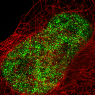
Figure 2: Active Motif's primary and fluorescent secondary antibodies in STED microscopy.HeLa cells were stained with alpha Tubulin mouse monoclonal antibody (Clone 5-B-1-2) (Catalog No. 39527), a biotinylated secondary goat anti-mouse antibody and BD Horizon V500-streptavidin conjugate. Histone H3 was stained with Histone H3 trimethyl Lys4 rabbit polyclonal antibody (Catalog No. 39159) and Chromeo 505 Goat anti-rabbit IgG (Catalog No. 15040) secondary antibody. The STED image is courtesy of Leica Microsystems, Germany.