Chariot™は活性のあるタンパク質・ペプチド・抗体などを2時間以内に哺乳細胞へ迅速かつ効率的に導入できる試薬です*。導入効率はおよそ60〜95%です。タンパク質導入後,導入したタンパク質などの作用を解析するために細胞をアッセイすることができます。その際,細胞固定の必要はありません。そのため,Chariotは阻害物質の導入,オルガネラの標識,ペプチドライブラリーのスクリーニングなど様々な用途に使用できます。
* ChariotはU.S.特許No.6,841,535を取得しています。研究目的用にのみ販売しています。他の目的でご使用の場合には,アクティブ・モティフ社までご連絡下さい。
| Name | Format | Cat No. | 価格 (税抜) | |
|---|---|---|---|---|
| Chariot™ | 25 rxns | 30025 | ¥82,000 | Buy |
| 100 rxns | 30100 | ¥188,000 | Buy | |
| β-Galactosidase Staining Kit | 75 rxns | 35001 | Discontinued | |
| Chariot™ Manual |
| β-Galactosidase Staining Kit Manual |
| Chariot™ Profile |
| Cell Biology Products Brochure |
| Chariot™ Citations |
Chariot™ の利点
- さまざまな種類のセルラインで使用できます (詳しくは User Data タブをご覧下さい)。
- 細胞固定の必要がなく, 生細胞でのタンパク質機能解析が可能です。
- in vivo1で使用できます。
- 2時間以内に60-95%の細胞内に導入されます。
- 融合タンパク質を発現させる必要がありません。
- 細胞毒性がなく,血清の有無に関わらず使用できます。
Chariot™の特長
現在,遺伝子導入にはカオチン脂質を用いた方法2, リン酸カルシウム法3, エレクトロポレーション法4, マイクロインジェクション法5 , ウィルスベクターを用いた方法6などの技術が使用されています。これらの方法は有用ですが,目的遺伝子の発現を解析するまでに,12-80時間待たなくてはなりません。また,これらの方法で一番問題になるのは細胞毒性です。細胞毒性があると,トランスフェクション後に観察される変化が,組換えタンパク質によるものか,もしくはトランスフェクション自体によるものかの判断が難しくなります。
Chariotはペプチドで,導入する高分子と非共有結合します。この複合体はタンパク質を安定化させるため,タンパク質は導入の間に分解されることなく機能が保たれます。7, 8. 培地にChariot/高分子複合体を加えると,すぐに細胞内に導入され,複合体は解離し,核移行シグナルをもChariotは核内へ移行し分解されます。解離した高分子は本来の活性を保っており,この高分子にシグナル配列がある場合には,標的細胞小器官へと運ばれます。またChariotはタンパク質,ペプチド,抗体を直接導入することにより,遺伝子発現に付随する転写- 翻訳プロセスを完全に省くため,時間を節約することができます。
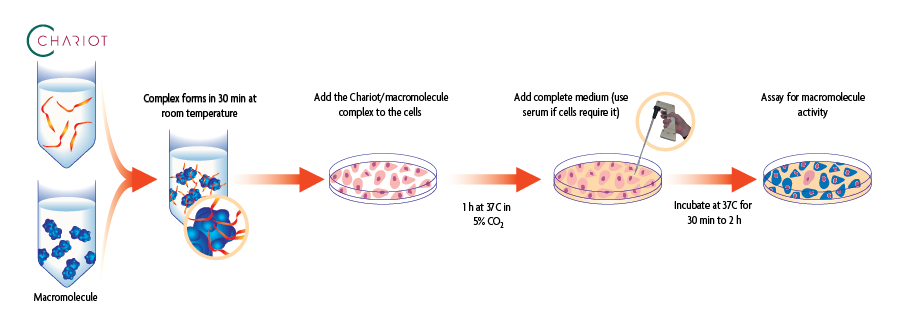
図1: Chariotを用いた目的高分子の細胞内への導入
Chariotの導入実績
Chariotは様々なセルラインへ効率的にタンパク質を導入できることが確認されています。 一般に分子量の大きいタンパク質を細胞内へ導入する場合, 導入には時間を要します。タンパク質の分子量が大きくなると, 導入効率が減少する可能性はありますが, 時間を長くすることで適切に導入されます。一方, Chariotは分子量が極端に小さいペプチドは導入できません。これはChariotペプチドと複合体を形成できないためです。そのためペプチドを使用する場合には少なくとも10アミノ酸以上を推奨しています。
Chariot™ 導入例
抗体
Chariotを用いてFITC標識, 抗Actin抗体および抗 Lamp-1抗体を導入した。これらのターゲットはそれぞれ, アクチンフィラメント (図 1) とエンドソーム (図2)である。
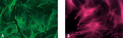
図1: 抗Actin抗体の導入
FITC標識, 抗Actin抗体(1/1000希釈)を用いてヒト繊維芽細胞 (HS-68) のアクチンフィラメントを標識した。抗体導入2時間後に, それぞれ細胞を固定 (A) したものと, 細胞を固定せず (B) に観察した。
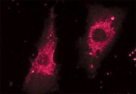
図2: 抗 Lamp-1抗体の導入
抗 Lamp-1抗体(1/500希釈)を用いてヒト繊維芽細胞 (HS-68) のエンドソームを標識した。抗体導入2時間後に, 細胞を固定せずに観察した。.
タンパク質
β-galactosidase は4個のサブユニットからなるタンパク質です。Chariotによるタンパク質導入効率を解析するため, β-galactosidase の 119 kDa サブユニットを用いた (図 3).
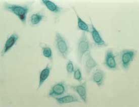
図3: HeLa細胞への β-galactosidase の導入
1 µg のβ-galactosidase 119 kDa サブユニットと Chariot™ Protein Delivery Reagent を混合し, 30 分間反応させ, HeLa 細胞に導入した。導入2時間後, 細胞を固定して β-Galactosidase Staining Kitを用いて染色した。
標的細胞小器官へ導入できます
非共有結合したChariotと高分子は細胞内へと導入されると解離します。, 解離した高分子は本来の活性を保っており,この高分子にシグナル配列がある場合には,標的細胞小器官へと運ばれます。これを示すために, 51-mer の核タンパク質(図 4) と 32-mer の細胞質ペプチドを用いた (図 5)。
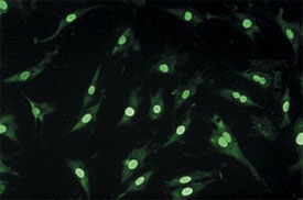
図4: 核タンパク質の導入
Chariot と蛍光標識した51-mer の核内タンパク質を 20:1 の割合で混合した後 HS-68 細胞へ導入した。導入90分後に, 細胞を固定せずに観察した。
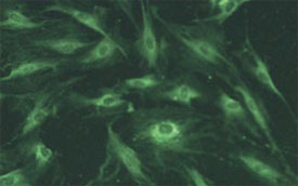
図 5: 細胞質ペプチドの導入
Chariot と蛍光標識した32-mer の細胞質ペプチドを 20:1 の割合で混合した後 3T3繊維芽細胞へ導入した。導入90分後に, 細胞を固定せずに観察した。C
参考文献
- Aoshiba, K. et al. (2003) Am. J. Resp. Cell & Mol. Biol. 28: 555-562.
- Felgner, P.L. et al. (1987) Proc. Natl. Acad. Sci. USA 84: 7413-7417.
- Graham, F.L. and van der Eb, A.J. (1973) Virology 52: 456-467.
- Neumann, E. et al. (1982) EMBO J. 7: 841-845.
- Capecchi, M.R. (1980) Cell 22: 479-488.
- Cepko, C.L. et al. (1984) Cell 37: 1053-1062.
- Morris, M. et al. (1999) J. Biol. Chem. 274 (35): 24941-24946.
- Morris, M. et al. (2001) Nature Biotech 19: 1173-1176.
Chariot’s ability to effectively deliver proteins, peptides and antibodies into a variety of cell types is well documented. To download a list of nearly 100 publications citing the use of Chariot, click here.
The following table is compiled from data furnished to us by our customers. This chart is by no means exhaustive.
| Cell line/model | Cell types | Macromolecule delivered | Transfection efficiency |
|---|---|---|---|
| 293 | Human embryonic kidney | GFP | 80% |
| 3T3-L1 | Mouse fibroblast | Antibody | 80% |
| A549 | Human lung carcinoma | Antibody | 95% |
| Arabidopsis | Primary plant protoplasts | Protein | Not provided |
| COS-7 | Monkey kidney | Protein, oligopeptide | 80% |
| CV-1 | Monkey kidney | Antibody | 55% |
| Embryonic cells | Primary mouse | Protein | 90% |
| Fetal Endothelial cells | Primary mouse | Protein | 70-80% |
| HCA2 | Human fibroblasts | Protein | 90% |
| HeLa | Human cervix carcinoma | Protein, peptide, antibody | 95% |
| HEK293 | Human kidney | Protein | 50% |
| HMSC | Primary human mesenchymal stem cells | Peptide | 80% |
| HS-68 | Human foreskin fibroblast | Protein, peptide, antibody | 95% |
| IMR90 | Human fibroblast | Protein, peptide | 80% |
| J774 | Mouse macrophage | Antibody | 90% |
| Jurkat | Human T-cell leukemia | Protein, peptide, antibody | 75% |
| MCF-7 | Human breast carcinoma | GFP | 70% |
| Mono Mac 6 | Primary human monocyte | Protein | 50% |
| Mouse hepatocytes | Primary liver | Protein | 95% |
| Mouse model (in vivo) | Alveolar wall tissue | Protein | 88% |
| Neural retina cells | Primary chicken | Protein | 80% |
| NIH/3T3 | Mouse embryo | Protein, peptide | 80% |
| NRK | Normal rat kidney | Antibody | 30-60% |
| Pancreatic cells | Primary human | Protein | 90% |
| PC-12 | Rat pheochromocytoma | Protein, peptide | 80% |
| Saos-2 | Human osteosarcoma cells | Antibody | 50-70% |
| SCC-61 | Primary human head + neck tumor | Peptide | 90% |
| Sensory neurons (DRG) | Primary chicken | Antibody, peptide | 80% |
| Sensory neurons (DRG) | Primary rat | Protein | 80% |
| Thyrocytes | Primary human | Protein | 90% |
| U373 | Human astrocytoma | Protein | 80% |
| U87mg | Human astroglioma | Antibody | 70% |
| WI-38 | Human lung fibroblast | Protein, peptide, antibody | 95% |
| WISH | Human placenta carcinoma | Protein | 30% |
Contents & Storage
Chariot is offered in 25- and 100-reaction kits, where a reaction is defined as the amount of reagents required to deliver the β-galactosidase positive control protein to cells in one well of a 6-well plate or in a 35 mm plate. The number of reactions you can perform will vary depending on the size plates used and the type of macromolecule(s) delivered. Each Chariot Kit contains sufficient Chariot Transfection Reagent, PBS, β-galactosidase positive control protein and sterile water for either 25 or 100 reactions. Store all reagents at -20°C. All reagents are guaranteed stable for 6 months.
Search our database of customer publications that have used our Chariot™ Protein Delivery Reagent.