The TCS STED (STimulated Emission Depletion) microscope from Leica Microsystems contains a pulsed 640 nm excitation laser and a 750 nm depletion laser that enable high-resolution microscopy with the red fluorescent dyes ATTO 647N and ATTO 655. The microscope can also be upgraded for use in dual color STED microscopy simply by integrating a second excitation laser of 531 nm. Active Motif’s fluorescent Chromeo™ 494 Dye and conjugated secondary antibodies meet the specifications of the second TCS STED laser combination and have been certified by Leica Microsystems for this application.
Use of Chromeo 494 and Active Motif’s ATTO (STED) secondary antibody conjugates enable you to perform co-localization studies at resolutions below the diffraction limit. This new method makes it possible to view the nuclear architecture or structures at synapses in more detail, which will help discovering and understanding sub-cellular structures and their role in cellular function.
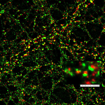
Figure 1: Pre- and post-synaptic marker proteins acquired by STED microscopy.
The localization of pre- and post-synaptic marker proteins visualized in the dendrites of nerve cells. The pre-synaptic protein Bassoon was stained by ATTO 647N (red), while the post-synaptic protein Homer was stained with Chromeo 494 (green). This STED image is courtesy of Dr. W. Zuschratter, IfN Magdeburg, Germany.
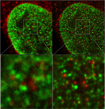
Figure 2: Nuclear structures visualized by dual color STED experiments.
The image on the left was prepared using a confocal microscope, while that on the right was produced using a STED microscope. The nuclear structures have been visualized with Chromeo 494 (green) and ATTO 647N (red). Images courtesy of Dr. L. Schermelleh, LMU Biozentrum Munich, Germany.
Chromeo 494 is available in a variety of attachment chemistries (reactive NHS-Ester, etc.) or in complete, optimized Fluorescent Antibody Labeling Kits, which make it easy to label primary antibodies and proteins.
To download the STED Microscopy Products Profile, please click here.