Histone H2A.XS139ph antibody (pAb)
| RRID: AB_2793161 |
| Aliases: Histone H2A.X phosphoserine 139 |
| 商品コード: 39117 | Format: 100 µg | Discontinued | |
| 商品コード: 39118 | Format: 10 µg | Discontinued | |
Applications


Application Notes
This product has been discontinued:
Please refer to our alternative product Catalog No. 91395
Published Applications
The following applications have been published using this antibody. Unless noted above, Active Motif may not have validated the antibody for use in these applications:
ICC/IF
ChIP-Seq
View publications that use Active Motif products & services here. Enter the product number(s) to see publications & applications that use this antibody.
Immunogen
This Histone H2AX phospho Ser139 antibody was raised against a peptide including phosphoserine 139 of histone H2AX.
Buffer
PBS containing 30% glycerol, 0.035% sodium azide. Sodium azide is highly toxic.
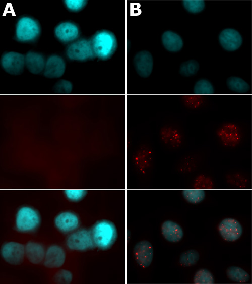
Histone H2AX phospho Ser139 antibody tested by immunofluorescence.
HeLa cells stained with Histone H2AX phospho Ser139 antibody (1:500 dilution) using MAX Stain™ Immunofluorescence Tools. The HeLa cells were blocked with MAXblock™ Blocking Medium and stained with Histone H2AX phospho Ser139 antibody.
Panel A: Untreated HeLa cells.
Panel B: Cells fixed and stained 90 minutes after 3 Gy ionizing radiation treatment.
Top images: Cells were stained with DAPI.
Middle images: Same cells stained with Histone H2AX phospho Ser139 antibody.
Bottom images: Merge of both images above.
Images were made using Zeiss Axiovision with equivalent acquisition settings for direct comparison. Note Panel B-middle image, which shows intense nuclear clustering of ionizing radiation-induced phosphorylation of Ser139 of H2AX. In contrast, Panel A-middle image shows no detectable phosphorylated H2AX.
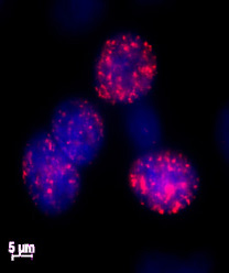
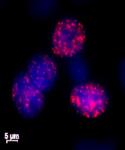
Histone H2AX phospho Ser139 antibody tested by immunofluorescence.
Etoposide-treated HeLa cells stained with Cat. No. 39117 (1:500 dilution, red) and counterstained with DAPI (blue).
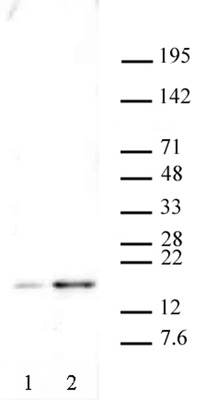
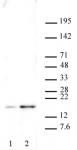
Histone H2AX phospho Ser139 antibody tested by Western blot.
Western blot: Nuclear extract of U2OS cells (20 µg per lane) probed with Histone H2AX phospho Ser139 polyclonal antibody (1:500 dilution).
Lane 1: untreated cells
Lane 2: cells treated with camptothecin
Storage
Some products may be shipped at room temperature. This will not affect their stability or performance. Avoid repeated freeze/thaw cycles by aliquoting items into single-use fractions for storage at -20°C for up to 2 years. Keep all reagents on ice when not in storage.
Guarantee
This product is guaranteed for 12 months from date of receipt.
This product is for research use only and is not for use in diagnostic procedures.
Application Key
- ChIP = Chromatin Immunoprecipitation;
- ChIP = ChIP Sequencing;
- CUT&RUN = Cleavage Under Targets and Release Using Nuclease;
- CUT&Tag = Cleavage Under Targets and Tagmentation;
- DB = Dot Blot;
- ELISA = Enzyme-linked Immunosorbent Assay;
- EMSA = Electrophoretic Mobility Shift Assay
- FC = Flow Cytometry;
- ICC = Immunocytochemistry;
- IF = Immunofluorescence;
- IHC = Immunohistochemistry;
- IP = Immunoprecipitation;
- MeDIP = Methyl-DNA Immunoprecipitation;
- NEU = Neutralizing;
- TIP-ChIP = Tagmented, Indexed, and Pooled Chromatin Immunoprecipitation;
- WB = Western Blotting